DDMFR Part A - purpose of assessment statement
Module 1 - Anatomy
The purpose of the examination is to assess whether those undertaking speciality training in Dental and Maxillofacial Radiology have an appropriate knowledge of the anatomy that underpins all radiological imaging including radiography, ultrasound, computed tomography (including cone beam CT), magnetic resonance imaging and sialography. Basic interpretation of chest radiography will also be expected.
Radiological anatomy is the cornerstone of clinical radiology for several reasons:
- A sufficient understanding is a prerequisite to performing any radiological study
- Radiologists need a thorough knowledge of normal radiological anatomy so
as to be able to recognise abnormalities - Radiologists need to be able to competently and accurately articulate the anatomical site of any abnormality to clinical colleagues
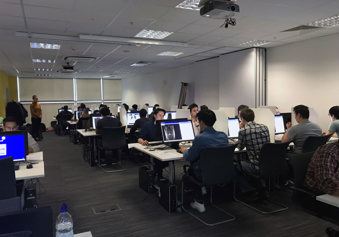
This module of the examination consists of a written paper and an anatomy image viewing component.
The written paper requires the candidate to answer 4 questions in 1 hour. Diagrams, where appropriate, are encouraged and answers may be presented as bullet points. The written paper assesses the understanding of anatomical relationships and the ability of the
candidate to communicate in writing and answer questions in a structured manner.
The anatomy image viewing component seeks to mimic what radiologists actually encounter in clinical practice and is therefore presented in electronic format. The examination consists of 100 questions, the majority of which are indicated by an arrow pointing to a specific anatomical structure. The question asked is typically “name the arrowed structure’’ with the answer recorded electronically. The reasoning behind this format is very simple; recognising a radiological anatomical structure and unprompted recall of its precise name is a key aspect of the everyday work of clinical radiologists. It is also vital to name the side of the structure where appropriate. Furthermore, doing so in a timely manner without routine recourse to reference material reflects real-life practice. The primacy of precision and clarity of communication about radiological anatomy is reflected in the way
the examination is constructed and marked. Examiners do take account of variable terms used for the same structure. In addition, the candidate will also be expected to be identify image faults. These are
included as the radiologist may be required to offer advice regarding image faults to radiographers and clinicians.
Module 2 - Radiological Sciences and Techniques
The purpose of this examination is to assess the candidates’ ability to explain and communicate principals of legislation, radiation physics and radiation protection applicable to dental radiography. It includes quality assurance and the use of selection criteria.
The examination will also cover the techniques available to investigate clinical conditions and candidates will be expected to base their recommendations on current guidelines, including best use of intra-oral dental radiography, Dental/Oral Panoramic Tomography (DPT/OPT), cephalometric radiography, extra-oral radiography of the facial bones /skull, Cone Beam CT, sialography, interventional sialography, MRI, conventional CT, ultrasound and nuclear medicine including PET.
It is recognised that Dental and Maxillofacial Radiologists may be involved in the teaching and training of dentists and the wider dental team in understanding and applying the principals of radiation protection, legislation (Ionising Radiations Regulations 2017 and
Ionising Radiation (Medical Exposure) Regulations 2017), radiation physics, quality assurance and selection criteria in relation to dental radiography.
This module of the examination consists of a written paper only.
The written paper requires candidates to answer 6 questions in 2 hours. Diagrams and bullet points may be used where appropriate. The written paper assesses the understanding of radiological sciences and techniques, including current legislation and the ability of the
candidate to communicate in writing and to answer the questions in a structured manner.
If a candidate is unsuccessful in one module of the examination, they are only required to re-sit that part of the examination.
Our exams
Find out more about our FRCR exams in clinical radiology and clinical oncology, and DDMFR exams in dental and maxillofacial radiology.
